Officers
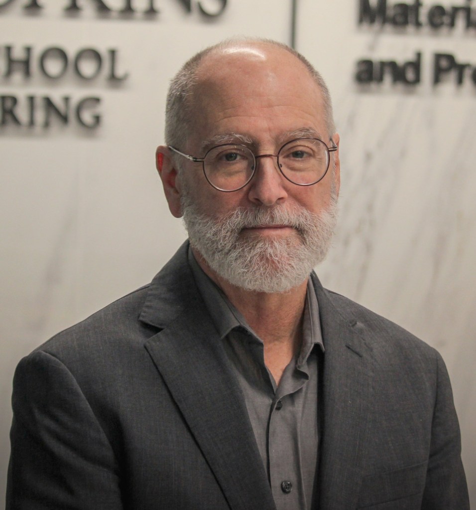
Ken Livi – President
Materials Characterization and Processing Facility, Johns Hopkins University
klivi@jhu.edu
Kenneth Livi is an associate research scientist in the Department of Materials Science and Engineering and director of operations for the Materials Characterization and Processing Facility. His research focuses on the characterization of inorganic and organic materials, biomaterials, earth materials, and surfaces of nanomaterials through electron microscopy.
He received a BS and MS from State University of New York at Stony Brook in 1980 and 1983, and a PhD at the Johns Hopkins University in 1995.
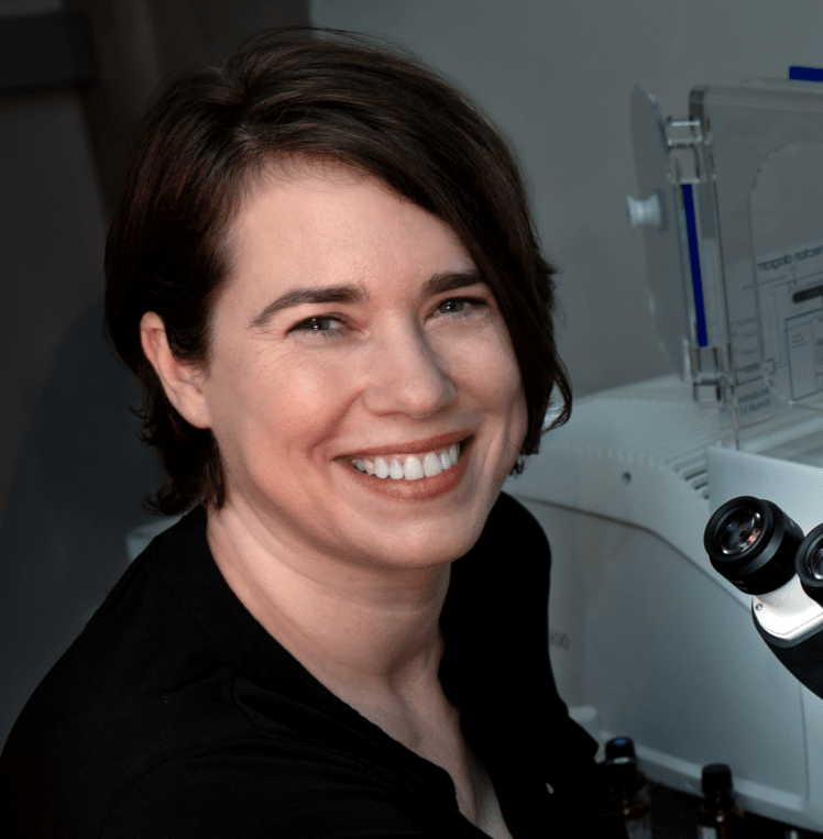
Tagide deCarvalho – President Elect
Keith R. Porter Imaging Facility, University of Maryland, Baltimore County
tagided@umbc.edu
Tagide deCarvalho is the director of the Keith. R. Porter Imaging Facility (KPIF) at UMBC where she oversees and performs optical and electron microscopy for on-campus faculty as well as external academic and industrial clients. She received a PhD in Behavior, Ecology, Evolution and Systematics from University of Maryland, College Park in 2009. Her postdoctoral work at the Carnegie Institution for Science, Department of Embryology involved extensive confocal microscopy, which set the stage for her career as a microscopist. She works with a wide variety of life and material science samples and has a special interest in imaging viral particles.
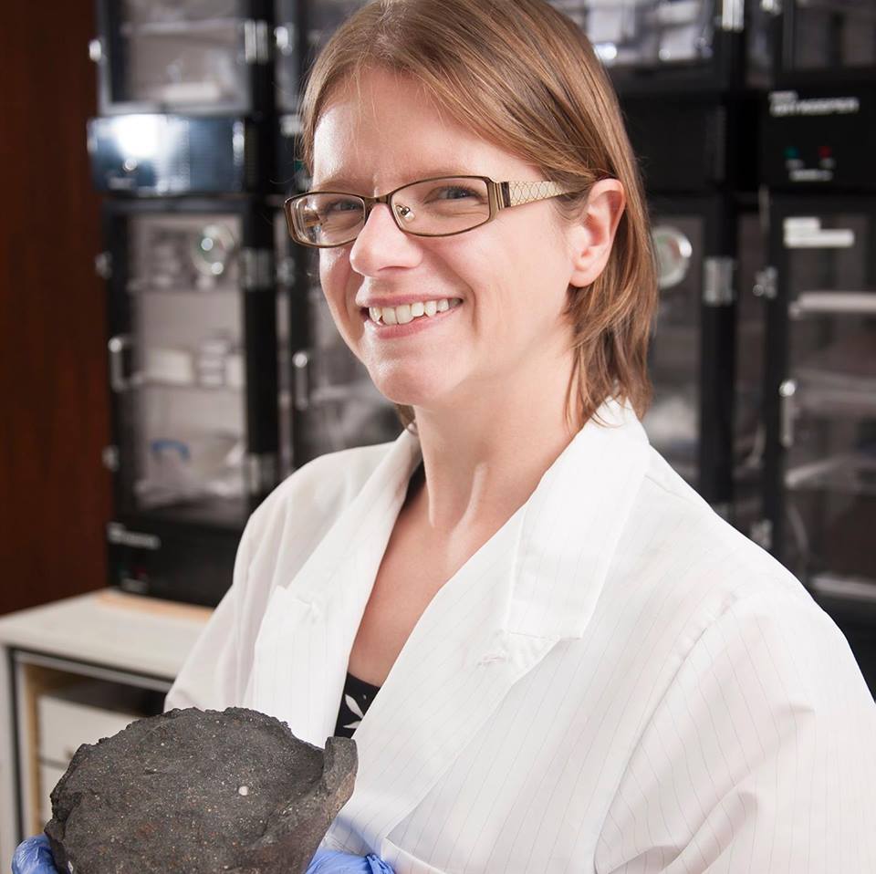
Emma Bullock – Treasurer
Earth and Planets Laboratory, Carnegie Institution for Science
ebullock@carnegiescience.edu
Emma Bullock is a Microbeam Specialist at the Earth and Planets Laboratory at the Carnegie Institution for Science. She operates Carnegie’s JEOL 8530 field emission electron microprobe, equipped with the xCLent cathodoluminescence detector, five WDS spectrometers, two EDS detectors, a cold trap for high precision carbon analysis and Probe for EPMA software. She also runs the Zeiss Auriga Scanning Electron Microscope (SEM) at Carnegie. She has authored and coauthored many papers in the fields of geology, planetary science and material science, and has expertise in imaging and quantitative analysis. Prior to joining Carnegie, she was a geochemist at the Smithsonian Institution.
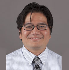
Thomas Lam – Secretary
Smithsonian Museum Conservation Institute
LamT@si.edu
Thomas Lam has a Ph.D. in Ceramics from Alfred University, NY. After earning his Ph.D., he completed postdoc at the National Institute of Standards and Technology. He is a physical scientist at the Museum Conservation Institute (MCI), where he applies his knowledge of material science and characterization skills (including scanning electron microscopy electron dispersive spectroscopy (SEM-EDS), cathodoluminescence (CL), X-ray fluorescence (XRF), and microfade testing) as part of the MCI technical studies team. The electron microscope Thomas performs research and technical studies with atMCI is a Hitachi S3700-N variable pressure SEM equipped with Bruker XFlash 6│60 EDS detector, Bruker XTrace (35 micron micro-XRF with a mapping substage), and Gatan ChromCL2 for CL.
Andrea Blake Brothers – Outreach Officer
American University/Hall of Science
brothers@american.edu
Light (PLM), electron (TEM & SEM), and dual beam (FIB/SEM) microscopy instrumentation to include sample preparation, imaging, and spectral analysis techniques for biological, pharmaceutical, and materials science (including semiconductor) applications.
Andrea Blake Brothers joined American University in January 2019 as Instrument Coordinator in the Chemistry Department. She has worked in both academia and industry for 40 years, utilizing various microscopy and microanalysis techniques in experimental design to help solve problems. Research experience includes positions with The University of South Carolina, School of Medicine; Georgetown and George Washington Universities; Biovail Pharmaceuticals; the Howard Hughes Medical Institute, Janelia Farm Research Campus, Micron Technology, and Tescan.
Ru-Ching Hsia – Council
Electron Microscopy Laboratory, Frederick National Laboratory for Cancer Research, NCI, NIH,
Ru-ching.hsia@nih.gov
Ru-ching Hsia has a Ph.D. in Microbiology and Immunology from Stanford University. She is the Principal Scientist and Head of the Electron Microscopy Laboratory (EML), at the Frederick National Laboratory-National Cancer Institute (NCI). Before joining EML, she was a Professor and Director of the Electron Microscopy Core Imaging Facility at the University of Maryland, Baltimore and in the Department of Embryology, Carnegie Institution for Science, Baltimore. Ru-ching has extensive experience in transmission and scanning electron microscopy, light microscopy, instrumentation, and sample processing for biomedical research. She was a Council member of the Microscopy Society of America and the Program Chair for the 2023 Microscopy & Microanalysis conference. Ru-ching is also the organizer and host of the bimonthly BioEMTalks webinar series.
Past Officers
2019
Ru-Ching Hsia – President
Robert Pope – President Elect
Emma Bullock – Treasurer
Kedar Narayan – Secretary
Joseph Mowery – Communications Officer
Thomas Lam – Outreach Officer
2018
Cam Robinson
Christine Brantner
Robert Pope
Ru-Ching Hsia
2017
Cam Robinson
Christine Brantner
Robert Pope
Ru-Ching Hsia
